
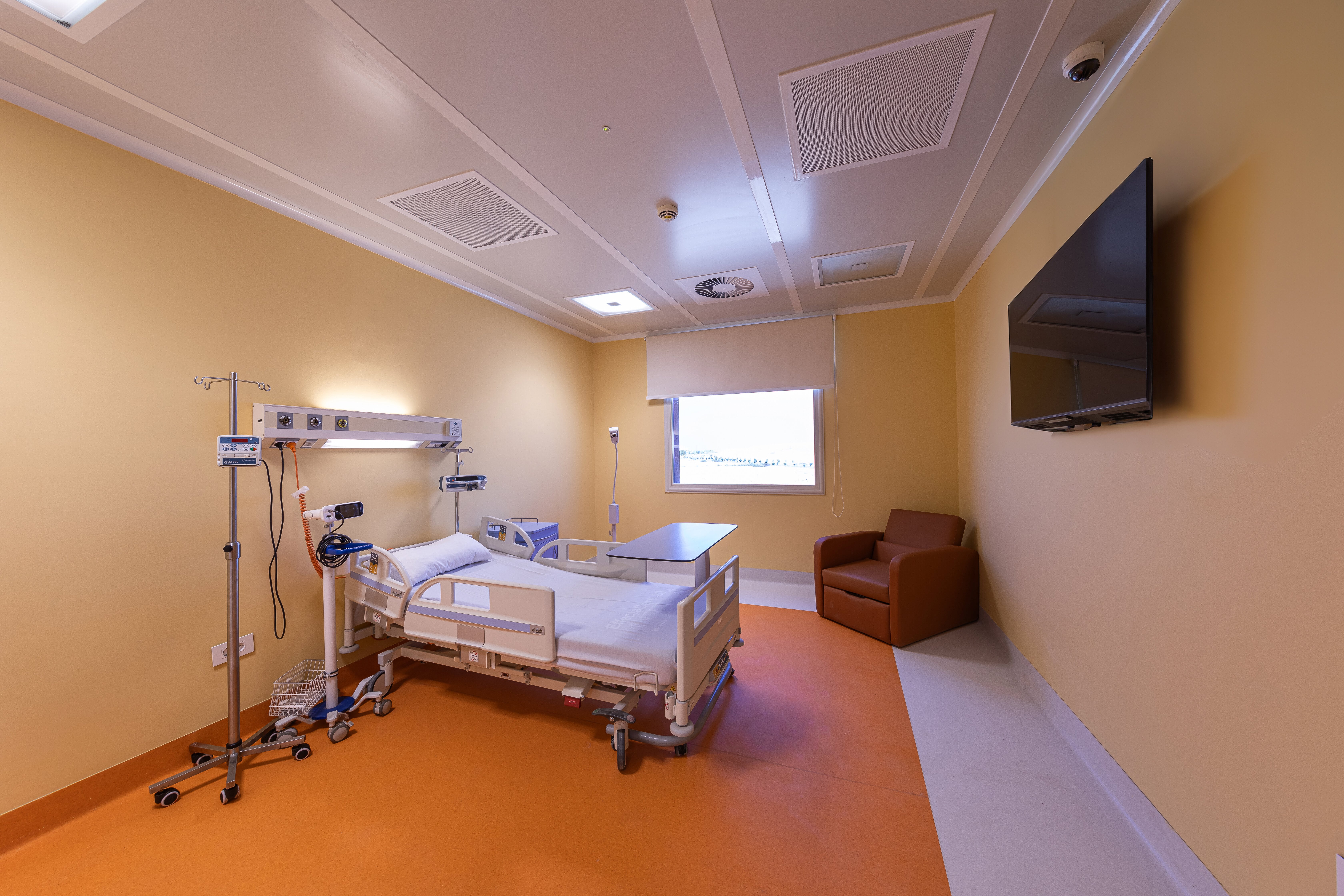
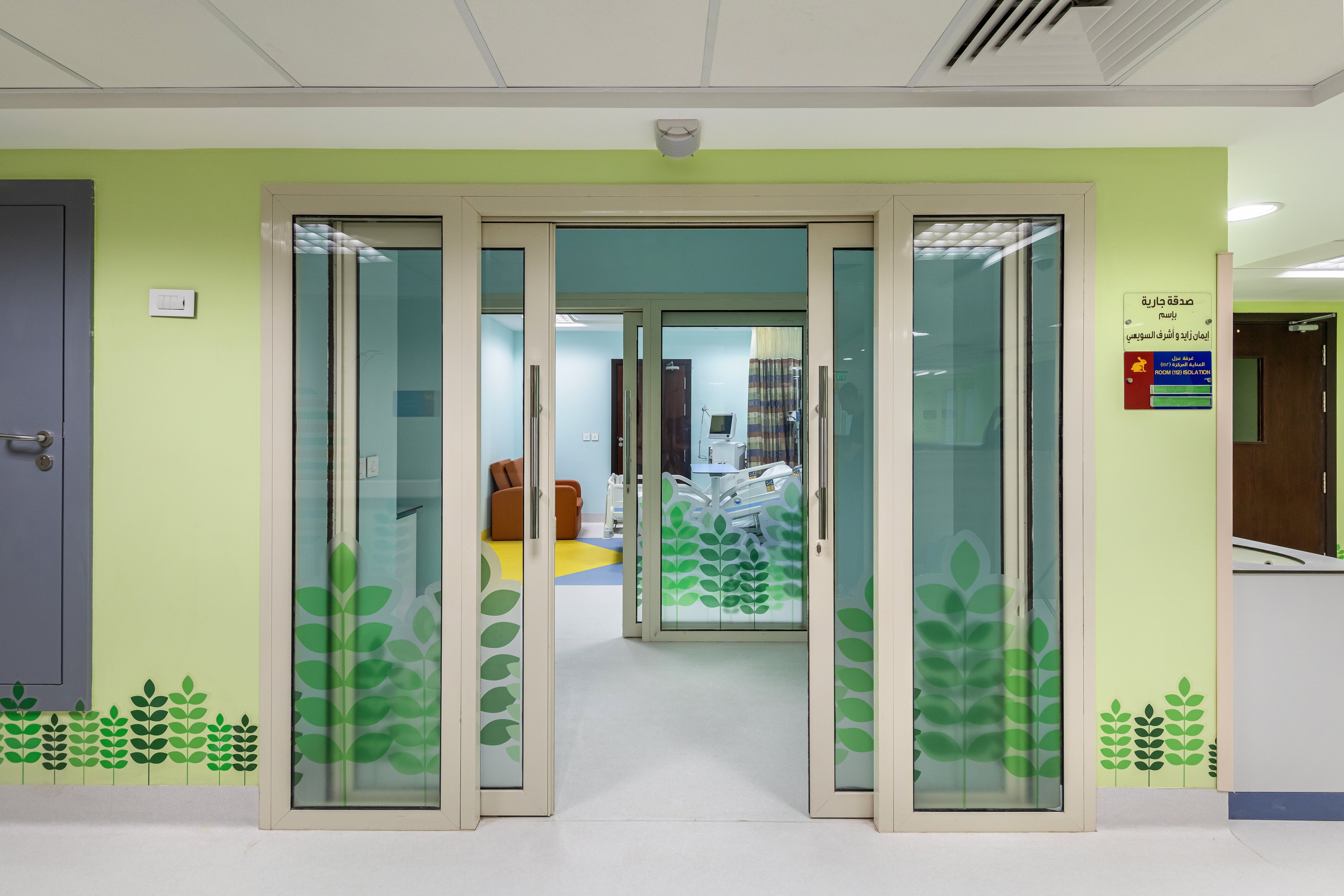
أنك ستكون تحت رعاية فريق عالمي من الأطباء والمتخصصين في مكان واحد. سوف يستمعون إلى قصتك، ويعملون معا لتشخيص حالتك وعلاجها بدقة حتى تتمكن من العودة إلى حياتك في أسرع وقت ممكن.
سيقوم فريق من الخبراء بوضع خطة العلاج الخاصة بك والاشراف على تنفيذها.
يضم فريق مستشفى شفاء الأورمان أفضل المتخصصين في كل مجال طبي تحتاجه، مع التركيز عليك. بغض النظر عن التحدي الصحي الذي تواجهه، يمكنك أن تكون واثقا من أنك تتلقى الرعاية الأكثر تقدما في مستشفى الشفاء الأورمان.
توفير راحة البال والأمل.
الحصول على التشخيص الصحيح في أسرع وقت يعني أنه يمكنك البدء في خطة العلاج الأكثر فعالية. نحن نعمل باستمرار على تطوير أدوات أفضل للتشخيص والعلاج، حتى تحصل على إجابات وخيارات وأمل حيث لم يكن هناك أي شيء من قبل.
نحن نركز على إنشاء خطة تشخيص وعلاج تناسبك.
سيقوم فريق من المتخصصين بتقييم حالتك بدقة وبعناية والعمل معا لوضع خطة مخصصة لتحقيق أهدافك. نهج فريقنا يجعل تجربتك أقل تعقيدا، حتى تتمكن من التركيز على التحسن.
CONSULTANTS RESPONSIBLE FOR THE PROGRAM:
1- Dr. Mohamed Mahmoud AbdElhakim
Consultant of radiotherapy at Shefa El Orman
- Specialization - Curriculum vitae
2- Dr. Emad shash
Consultant of medical oncology at Shefa El Orman
- Specialization - Curriculum vitae
3- Dr. Tarek M Hashem
Consultant of surgical oncology at Shefa El Orman
- Specialization - Curriculum vitae
4- Dr. Mohamed Adly El Ramely
Consultant of anesthesiology at Shefa El Orman
- Specialization - Curriculum vitae
Information for the patients:
- Breast cancer is a disease in which abnormal breast cells grow out of control and form a mass called a tumor. The tumor can grow to invade and destroy healthy body tissue. In time, cancer cells can break away and spread to other parts of the body. When cancer spreads, it's called metastatic cancer.
Incidence
- Breast cancer is the most common cancer diagnosed in women.
- Approximately 99% of breast cancers occur in women and 0.5–1% of breast cancers occur in men.
- Breast cancer survival rates have been increasing. Much of this is due to the widespread support for breast cancer awareness.
- Advances in breast cancer screening allow healthcare professionals to diagnose breast cancer earlier. Finding the cancer earlier makes it much more likely that the cancer can be cured.
CONSULTANTS RESPONSIBLE FOR THE PROGRAM:
1- Dr. Ayman Abdel Wahab Amin
Consultant of gastro enterology surgery at Shefa El Orman
- Specialization - Curriculum vitae
2- Dr. Mohamed Mahmoud AbdElhakim
Consultant of radiotherapy at Shefa El Orman
- Specialization - Curriculum vitae
3- Dr. Noha Rashad
Consultant of medical oncology at Shefa El Orman
- Specialization - Curriculum vitae
4- Dr. Mohamed Adly El Ramely
Consultant of anesthesiology at Shefa El Orman
- Specialization - Curriculum vitae
5- Dr. Shady El Sebaey
Consultant of intervention radiology at Shefa El Orman
- Specialization - Curriculum vitae
CONSULTANTS RESPONSIBLE FOR THE PROGRAM:
1- Dr. Ziyad Samir Sayed
Consultant of gynecological surgery at Shefa El Orman
- Specialization - Curriculum vitae
2- Dr. Mohamed Mahmoud AbdElhakim
Consultant of radiotherapy at Shefa El Orman
- Specialization - Curriculum vitae
3- Dr. Mariam Hussin
Consultant of clinical oncology at Shefa El Orman
- Specialization - Curriculum vitae
4- Dr. Mohamed Adly El Ramely
Consultant of anesthesiology at Shefa El Orman
Specialization - Curriculum vitae
CONSULTANTS RESPONSIBLE FOR THE PROGRAM:
5- Dr. Mohamed Mahmoud AbdElhakim
Consultant of radiotherapy at Shefa El Orman
- Specialization - Curriculum vitae
6- Dr. Osama Abdel Mohaimen
Consultant of medical oncology and hematology at Shefa El Orman
- Specialization - Curriculum vitae
CONSULTANTS RESPONSIBLE FOR THE PROGRAM:
1- Dr. Ayman Abdel Wahab Amin
Consultant of head and neck surgery at Shefa El Orman
- Specialization - Curriculum vitae
2- Dr. Mohamed Mahmoud AbdElhakim
Consultant of radiotherapy at Shefa El Orman
- Specialization - Curriculum vitae
3- Dr. Mariam Hussin
Consultant of clinical oncology at Shefa El Orman
- Specialization - Curriculum vitae
4- Dr. Mohamed Adly El Ramely
Consultant of anesthesiology at Shefa El Orman
- Specialization - Curriculum vitae
CONSULTANTS RESPONSIBLE FOR THE PROGRAM:
9- Dr. Shady El Sebaey
Consultant of intervention radiology at Shefa El Orman
- Specialization - Curriculum vitae
10- Dr. Mohamed Adly El Ramely
Consultant of anesthesiology at Shefa El Orman
- Specialization - Curriculum vitae
Including:
CONSULTANTS RESPONSIBLE FOR THE PROGRAM:
1-
Dr. Galal Rafik Galy
Consultant of thoracic surgery at Shefa El Orman
- Specialization - Curriculum vitae
2-
Dr. Amr Mahmoud El Demery
Consultant of thoracic surgery at Shefa El Orman
- Specialization - Curriculum vitae
3-
Dr. Mohamed Mahmoud AbdElhakim
Consultant of radiotherapy at Shefa El Orman
- Specialization - Curriculum vitae
4-
Dr. Maha Yahia
Consultant of medical oncology at Shefa El Orman
- Specialization - Curriculum vitae
5-
Dr. Mohamed Adly El Ramely
Consultant of anesthesiology at Shefa El Orman
- Specialization - Curriculum vitae
6-
Dr. Shady El Sebaey
Consultant of intervention radiology at Shefa El Orman
- Specialization - Curriculum vitae
Information for the patients: Lung cancer begins when epithelial cells, which form the inside lining of the lungs, grow rapidly and uncontrollably. Often, these cells develop into a mass called a tumor. A malignant or cancerous tumor can stay in one place or spread to other parts of the body. Early lung cancer often has no symptoms and can only be detected by medical imaging. As the cancer progresses, most people experience nonspecific respiratory problems: coughing, shortness of breath, or chest pain. Other symptoms depend on the location and size of the tumor. Incidence: • Lung cancer is the third most common cancer type worldwide.
• The average age of diagnosis is 65-70 years old.
• Men are twice as likely to be diagnosed with lung cancer, which largely reflects differences in tobacco consumption.
• Lung cancer is the leading cause of cancer-related deaths worldwide, accounting for the highest mortality rates among both men and women.
• Smoking is the leading cause of lung cancer, responsible for approximately 85% of all cases.
CONSULTANTS RESPONSIBLE FOR THE PROGRAM:
11- Dr. Mohamed Ahmed Ebrahim
Consultant of sarcoma surgery at Shefa El Orman
- Specialization - Curriculum vitae
12- Dr. Mohamed Mahmoud AbdElhakim
Consultant of radiotherapy at Shefa El Orman
- Specialization - Curriculum vitae
13- Dr. Maha Yahia
Consultant of medical oncology at Shefa El Orman
- Specialization - Curriculum vitae
14- Dr. Mohamed Adly El Ramely
Consultant of anesthesiology at Shefa El Orman
- Specialization - Curriculum vitae
Information for the patients:
A sarcoma is a rare type of malignant (cancerous) tumor that develops in bone and connective tissue, such as fat, muscle, blood vessels, nerves and the tissue that surrounds bones and joints. Symptoms depend on the tumor’s size and location. Treatments include surgery, radiation, chemotherapy, targeted therapy and immunotherapy.
Sarcomas form when immature bone or soft tissue cells have changes to their DNA, and they develop into cancer cells that grow in an unregulated way. They may eventually form a mass or tumor that can invade nearby healthy tissues. Left untreated, the cancer can travel via your bloodstream or lymphatic system from the primary location where it formed to other organs (metastasis). Metastasized cancer is challenging to treat.
Sarcomas making up only 1% of all adult cancer diagnoses and about 15% of childhood cancer diagnoses. Approximately 16,000 sarcomas are diagnosed each year (around 4,000 bone sarcomas and approximately 13,000 soft tissue sarcomas). Bone sarcomas are more common among children while soft tissue sarcomas are more common in adults.
Sarcomas are categorized as soft tissue or bone sarcomas, depending on where they develop in the body.
Soft tissue sarcomas originate in the soft tissues of the body and are most commonly found in the arms, legs, chest or abdomen. Unlike bone sarcomas, most soft tissue sarcomas occur in adults. Certain types of sarcoma, such as rhabdomyosarcoma, are found mostly in children.
Types of soft tissue sarcomas include:
Primary bone sarcoma is cancer that starts in the bone. More than one-third of bone sarcomas are diagnosed in people younger than 35 years old. Many are diagnosed in children.
Types of primary bone sarcoma include:
Symptoms:
Symptoms vary depending on the tumor’s location. For example, some sarcomas may not cause noticeable symptoms in the early stages. Some sarcomas may feel like a painless lump under your skin, while others don’t cause pain until they grow large enough to press on an organ.
Other sarcomas can cause long-lasting bone pain or swelling in your arm or leg that worsens at night. These changes may limit your movement.
Symptoms may include:
Risk factors:
Most sarcomas do not have a known cause, although there are several factors that could increase a person’s risk of developing a sarcoma. The most common sarcoma risk factors include the following:
Inherited syndromes. Some syndromes that increase the risk of cancer can be passed from parents to children. Examples of syndromes that increase the risk of sarcoma include familial retinoblastoma and neurofibromatosis type 1.
Radiation therapy for cancer. Radiation treatment for cancer increases the risk of developing a sarcoma later.
Chronic swelling (lymphedema). Lymphedema is swelling caused by a backup of lymph fluid that occurs when the lymphatic system is blocked or damaged. It increases the risk of a type of sarcoma called angiosarcoma.
Exposure to chemicals. Certain chemicals, such as some industrial chemicals and herbicides, can increase the risk of sarcoma that affects the liver.
Exposure to viruses. The virus called human herpesvirus 8 can increase the risk of a type of sarcoma called Kaposi's sarcoma in people with weakened immune systems.
Diagnosis :
Your healthcare provider will begin with a thorough history and physical examination. They may perform special tests on a tissue sample obtained from a biopsy to diagnose the exact type of sarcoma you have.
They may use any of the following tests when making a diagnosis:
Stages of sarcoma:
Cancer staging is used to better understand how serious a sarcoma is and what treatments may work best. Providers classify most sarcoma based on the TNM classification system:
Providers use this information to stage your sarcoma. They assign a number (1 through 4) to your stage. The higher the number, the more the cancer has grown locally or spread throughout your body.
Treatment:
Sarcoma is treated with a combination of chemotherapy, radiation therapy and surgery. Reconstruction of the surgical area typically takes place at the same time the tumor is removed.
Your treatment will depend on various factors, including:
For primary tumors, radiation is used in conjunction with surgery (either before or after) to reduce the risk of tumor recurrence.
Patients with isolated metastasis might be treated with radiation in conjunction with chemotherapy as well as stereotactic radiosurgery. Radiation is an essential part of treating most high-grade tumors.
Surgery
Your provider will work to remove all cancer cells while sparing as much healthy tissue as possible. They’ll perform a wide local excision, while leaving a margin of healthy tissue at the edges. This approach removes the tumor without leaving any microscopic disease.
Rebuilding the affected area is also essential. Limb salvage (saving/preserving an arm or a leg) may involve removal of the tumor and, if needed, metal replacements for joint reconstruction. It may include replacing the bone removed during surgery with bone taken from another part of your body or elsewhere as needed. In some instances, the safest and best option is to amputate the affected limb, which can be an especially difficult decision.
Radiation therapy
Radiation therapy uses high-energy X-rays to kill cancer cells while minimizing damage to healthy cells. Radiation therapy may be internal (placed in your body) or external (delivered by a machine outside your body). Radiation uses advanced techniques so that radiation is directed toward the cancer while sparing healthy areas.
Internal radiation, also called brachytherapy, can be used to treat sarcoma that hasn’t spread. Brachytherapy can be delivered in two ways: intra-operative radiotherapy (during surgery) and interstitial brachytherapy, which is delivered through a series of catheters (plastic tubes) after surgery.
Radiation can also treat sarcoma that's spread (metastatic disease).
Chemotherapy
Chemotherapy uses drugs that kill or slow the growth of rapidly multiplying cancer cells. These medications are given intravenously (through a needle into a blood vessel) or orally. You may receive chemotherapy before surgery to shrink the tumor or after surgery. In some cases, chemotherapy is given with radiation.
Targeted therapy
Targeted therapy attacks weaknesses in cancer cells without harming healthy cells in the process. Targeted therapy only works on certain types of cancer cells with the weaknesses it’s designed to target. Your doctor may have your sarcoma cells tested to see if they are likely to respond to targeted therapy drugs.
Your provider may recommend targeted therapy as a standalone treatment or in combination with other treatments.
Immunotherapy
Immunotherapy is a drug treatment that uses your immune system to fight cancer. Your body's disease-fighting immune system may not attack your cancer because the cancer cells produce proteins that blind the immune system cells. Immunotherapy drugs work by interfering with that process.
Ablation therapy
Ablation therapy treatments destroy cancer cells by applying electricity to heat the cells, very cold liquid to freeze the cells or high-frequency ultrasound waves to damage the cells.
Palliative care
Palliative care is specialized medical care for people with serious illnesses, including cancer. Palliative care professionals include doctors, nurses, social workers and nutritionists, among others. They can help with symptom relief and help you manage treatment side effects. Your palliative care team can help you navigate your cancer diagnosis, ensuring your quality of life is always a top priority.
Sarcoma care at Shefa-El Orman Hospital:
Shefa-El Orman doctors are respected for their expertise in diagnosing and treating sarcoma. At Shefa-El Orman you can expect compassionate, comprehensive care that's focused on your individual needs.
At Shefa-El Orman adult and pediatric medical oncologists, radiation oncologists, orthopedic surgeons that specialize in sarcoma treatment, pathologists, and specialists in physical medicine and rehabilitation work as a multidisciplinary team to care for people with sarcoma. Other professionals are included as needed.
Shefa-El Orman doctors will work with you to review all of your treatment options and choose the treatment that best suits your needs and goals.
The range of treatments offered to people with sarcoma includes:
CONSULTANTS RESPONSIBLE FOR THE PROGRAM:
15-
Dr. Ahmed Abdel Bary
Consultant of urology surgery at Shefa El Orman
- Specialization - Curriculum vitae
16-
Dr. Mohamed Mahmoud AbdElhakim
Consultant of radiotherapy at Shefa El Orman
- Specialization - Curriculum vitae
17-
Dr. Mariam Hussin
Consultant of clinical oncology at Shefa El Orman
- Specialization - Curriculum vitae
18-
Dr. Mohamed Adly El Ramely
Consultant of anesthesiology at Shefa El Orman
- Specialization - Curriculum vitae
Glioblastoma multiforme Brain cancer is a malignant tumor of the cells of the brain (this implies the primary occurrence of a tumor in the brain and not cancer that has metastasized to the brain from other organs). In brain cancer, normal brain cells transform into cancerous ones. In a healthy body, cells appear in the right amount and die off at a certain time, giving the space for the new ones. Cancer cells begin to grow uncontrollably, without dying off. Their accumulation forms a brain tumor. Gliomas are the most common types of brain tumors. There are 4 degrees of malignancy of such formations, based on the type of cells of the nervous tissue. Glioblastoma is the most malignant type of brain tumor, characterized by the highest rate of growth and the absence of distinguishable borders. Thus, glioblastoma belongs to brain tumors of the highest degree of malignancy, which represent the greatest difficulties for medical science in the matter of treatment. The exact causes of brain cancer have not been established. It may be associated with a hereditary predisposition, as well as with the impact of dangerous working conditions.
- Symptoms:
The main warning signs for brain tumors are: • Frequent nausea and vomiting
• Headache in the morning or headache that goes away after vomiting
• Visual impairment, hearing impairment, problems with speech
• Loss of balance and unstable gait
• The weakness of one side of the body
• Unusual sleepiness or changes in daily activity levels
• Radical changes in personality or behavior
- Diagnosis:
When carrying out computed tomography, a layered image of brain tissue is obtained. Thus, CT allows assessing the size of brain tumors and the exact location of the neoplasms. MRI is a more detailed visualization of brain tissue, in which even the smallest changes in the anatomical structure of the brain are visible. Using functional magnetic resonance imaging, the doctor evaluates blood circulation in the brain tumors and brain tissues. Magnetic resonance spectroscopy is a research technique in which the metabolism in the tissues of brain tumors is investigated. Magnetic resonance angiography reflects the relationship between the brain tumor and large arterial and venous vessels.
Treatment:
Currently, there are three types of treatment for glioblastoma beyond stage one-three. These types are surgery, radiation therapy, and chemotherapy. At the present stage, progress has been made in each of the types of treatments, but their combination gives the best results.
• Surgery.
With the information on the exact localization of the brain tumor based on MRI results, the neurosurgeon can remove a tumor without subsequent side effects. In some cases, when the brain tumor is located close to the motor or speech centers, the surgery is performed under local anesthesia, which allows patients to be conscious and allows doctors to control the speech function. After the removal of glioma, most patients have an improvement in neurological symptoms. Postoperative mortality, even for glioblastoma, reaches 1%, and the worsening of the condition is seen in up to 5% of cases. The best results are achieved after radical resection (compared to partial tumor resection or biopsy). The development of innovative methods of diagnosis and treatments allows the surgeon to successfully perform the radical removal of glioblastomas in most cases of brain tumors of such types. If the tumor is located close to the functionally important cortical structures, the surgeon should consider performing functional MRI before surgery. Using stereotactic navigation and intraoperative monitoring, an attempt should be made to remove the entire brain tumor. Time spent on carefully performed resection will be rewarded with increased survival and quality of life for patients. If the brain tumor cannot be removed, a stereotactic biopsy should be performed. Patients with a metastasized type of brain cancer should receive radiation therapy followed by chemotherapy. If the disease recurs, resection of the tumor should be considered, followed by second-line chemotherapy, experimental therapy, or participation in the clinical trials.
• Radiation therapy.
Brain tumors often recur in their previous area. That is why total radiation therapy is often performed after the surgery in order to prevent the relapse. Another type of irradiation, the localized radiation therapy, is aimed precisely at tumor cells in the brain. The development of neuroimaging techniques allows the radiologist to reduce the complications of radiation therapy by targeting the brain tumor without affecting the healthy brain structures. Therefore, it is better to use both local radiation therapy and total radiation therapy for the whole brain. Even with these precautions, delayed damage to healthy brain tissue remains a significant long-term problem. At the present stage, stereotactic surgery is the most effective and safe method to treat brain tumors. This method can be considered an alternative to the traditional surgery. This allows a large dose of radiation to be delivered to the brain tumor without an additional radiation load on healthy tissues. Modern computer technologies in imaging make it possible to carry out this type of surgery accurately. None of the currently existing surgical systems can excise the brain tumor with the same precision. The effect of radiation therapy for glioblastoma can be assessed only in several months after the procedure. The tumor is not destroyed, but it stops growing and decreases in size. Stereotactic surgery is applied in the treatment of small brain tumors that do not exceed three centimeters in diameter and have clear boundaries. Stereotactic surgery is not suitable for rapidly growing brain tumors.
• Chemotherapy.
Cane be used in combination with radiotherapy and In the stage 4 glioblastoma treatment, chemotherapy should be considered the optimal treatment, since these tumors penetrate deep into the normal brain and tumor cells are often found at a distance of up to four centimeters from the primary focus. Chemotherapy drugs cross the blood-brain barrier and can reach every tumor cell in the central nervous system. Glioblastomas are heterogeneous. Genetic and biochemical abnormalities that have been identified in glioblastomas generally only occur in certain cells. Not all glioblastoma cells have the same histochemical features, and this must be considered when choosing the treatment of glioblastoma multiforme grade 4
مؤسسة شفا الأورمان المُشهره بوزارة التضامن الاجتماعي برقم 5859 لسنة 2016
جميع الحقوق محفوظة مستشفي شفاء الأورمان - صعيد بلا سرطان © 2024